Introducing Advanced Imaging for Pets: What Makes HDVI Special?
At Sunstone Veterinary Specialists, we are thrilled to introduce a groundbreaking imaging technology to our community: HDVI for pets. This advanced tool revolutionizes how we diagnose and treat our furry companions by offering a more detailed and precise look inside their bodies.
What is HDVI?
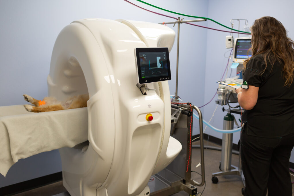
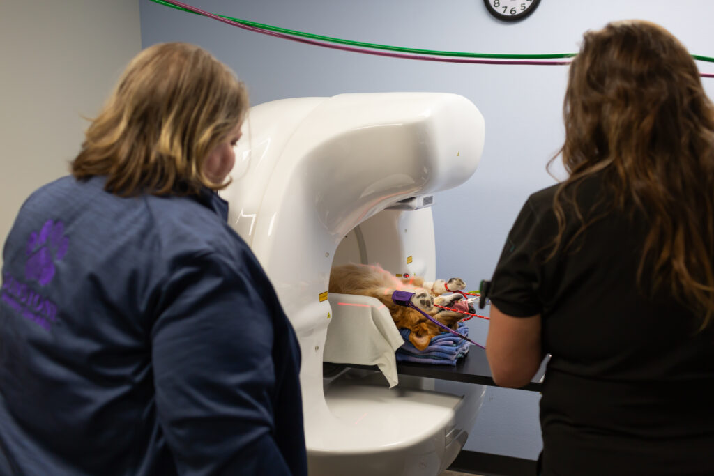
HDVI, or high-definition volumetric imaging, is a state-of-the-art diagnostic tool that takes veterinary imaging to the next level. While traditional CT (Computed Tomography) scans are widely known for their ability to create cross-sectional images of the body, HDVI goes a step further. Instead of generating “slices” of the body, HDVI collects true 3D images by seamlessly rotating 360 degrees around your pet.
Our HDVI system, the Vimago GT30, captures 720 images within a 6-inch section of the body in just 24 seconds. These images are combined into a single 3D “voxel,” producing incredibly detailed and accurate visuals. Unlike traditional CT scans, which rely on computers to estimate what lies between slices, HDVI creates a complete, true-to-life image.
How Does HDVI Work?
To ensure clear images, pets must stay completely still during the 24-second scan. Since this can be challenging for wiggly tails and twitchy whiskers, we use anesthesia to keep them calm and comfortable. In some cases, a contrast dye is injected to highlight specific areas, such as blood vessels or inflamed tissues, providing even more detail for our veterinary radiologists.
The process involves:
- A pre-contrast scan to capture the initial image.
- Injection of contrast material, either intravenously or into a specific area of interest.
- A post-contrast scan to enhance visualization of affected areas.
These images are then sent to Peregrine Radiology, specialists trained to analyze HDVI studies.
Why Choose HDVI for Your Pet?
HDVI offers several advantages over traditional diagnostic tools, including:
- Enhanced Precision: The 3D images are more detailed than traditional CT scans, helping us identify issues that might otherwise be missed.
- Non-Invasive: HDVI doesn’t harm tissues, making it a safe option for pets.
- Improved Pre-Surgical Planning: The detailed images allow surgeons to plan procedures like correcting liver shunts or repairing joints with greater accuracy.
Common Uses for HDVI
HDVI for pets is not typically the first diagnostic tool we use, as physical exams, x-rays, and blood tests often provide sufficient information. However, HDVI is invaluable for more complex cases, including:
- Diagnosing conditions in the chest and lungs, where tiny abnormalities may be invisible on x-rays.
- Preparing for surgeries, such as correcting liver shunts or evaluating malformed joints.
- Combining with other procedures, like rhinoscopy, to investigate the sinuses or collect tissue samples.
A New Standard in Veterinary Imaging
HDVI is a game-changer for veterinary diagnostics in the Portland Metro area. Its ability to provide clear, precise, and detailed images helps us deliver the best possible care for your pets. Whether it’s planning a surgery or diagnosing a complex condition, HDVI for pets offers a new level of insight and confidence.
If you have questions about HDVI or think it might be right for your pet, contact us at Sunstone Veterinary Specialists. Together, we can ensure your furry family member receives the advanced care they deserve.